


Neuroimaging and Neurostimulation Techniques: The Benefits & Limits of Each
From fMRI to TMS, discover the different types of neurofunctional technologies and learn about a study from the University of Washington that combines them to create something groundbreaking

The human brain is made of billions of neurons, and these neurons are constantly “talking” to each other by sending out electrical signals that produce tiny electric fields (about 40 microvolts). These tiny electric fields accumulate to the point that they become noticeable and are able to be detected by neuroimaging machines. Different activities produce different changes in electric and magnetic fields across the brain due to different levels and dynamics of neural communication. And from this, maps of the brain are ultimately created after a lot of complex data processing is done by computers that compile and analyze information from the accumulated electric fields. This data highlights what areas of the brain are active and when they’re active during different activities, providing doctors and researchers with insights into variable brain activity with differing levels of spatial and temporal resolution.
Read this article to find out more about these incredible neuroimaging and neurostimulation technologies, as well as an innovative telepathic study conducted by researchers at the University of Washington that uses a couple of them to produce something you might only expect to see in science fiction.
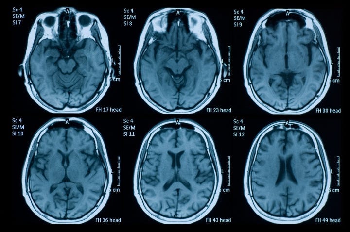
Spatial Resolution — fMRI
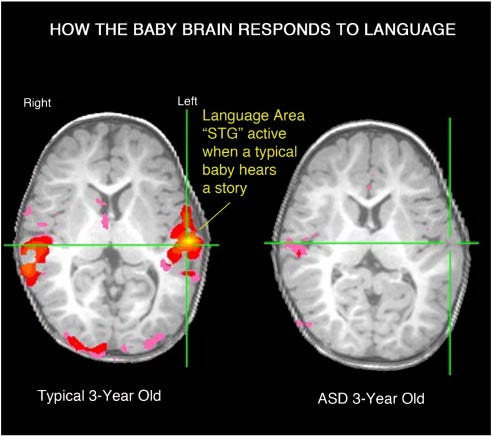
Through spatial resolution, you can determine which parts of the brain are active during a task: the where. And typically this is carried out with an fMRI (functional magnetic resonance imaging) machine, maps of the brain are created after a lot of complex data processing by detecting tracks of oxygenated blood flow through the brain. Areas of high oxygenated blood flow are associated with high neural activity. Here’s what an fMRI looks like in a typical 3-year old vs. a 3-year old with autism spectrum disorder (ASD), showing the language area (superior temporal gyrus, or STG) that lights up in response to hearing a story and the absence of activity in that area of the brain in babies with ASD:
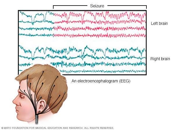
Temporal Resolution — EEG
Temporal resolution, which is mainly provided by electroencephalography scans, shows when the brain is active. It does so by recording electrical activity in the brain using electrodes placed directly on the scalp. This allows for causal inferences to be made; for instance, when you perform a particular task, imaging for temporal resolution allows researchers to determine specifically when certain parts of the brain light up. As a result, it is possible to infer a causal relationship between performing a task and what parts of the brain show activity when doing that task. However, unlike fMRI, EEG provides poor spatial resolution (and only shows surface-level activity; not 3D).
PET: Positron Emission Tomography
PET scans create images of the brain by detecting radioactively-labeled substances like water and glucose. While it is a commonly used neuroimaging technique, like fMRI, it does not really track neural activity–only blood flow / specific substances — producing a “picture” of the brain every 2 seconds. Because of this, it has very poor temporal resolution and is thus primarily correlational — no causal inference can be made (again EEG would be an example of CAUSAL). Nevertheless, PET scans are often used to diagnose brain disorders such as epilepsy, Alzheimer’s, and Parkinson’s by detecting patterns of abnormal neurological activity.
Electrical and Magnetic Stimulation in Brain Areas
Transcranial Magnetic Stimulation (TMS)
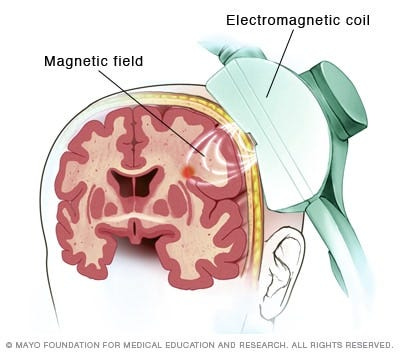
In TMS, electromagnetic coils are held against the scalp to activate neurons directly underneath the site of stimulation, and are also used to study neural circuitry responsible for a wide range of functions. It is a great method to create reversible, “virtual” lesions that allow you to create “visual patients” in the lab. There are also therapeutic applications of this type of neurostimulation, as one form of TMS called rTMS (repetitive transcranial magnetic stimulation) can be helpful in treating depression.
The Pros and Cons of TMS
Pros
Pinpoint a function to a particular region
Provide causal evidence (non-correlational)
Cons
Difficult to localize damage precisely
Binary manipulation (working/non-working)
Locality assumption (no network effects)
Optogenetics
A new technique to manipulate the activity of specific subsets of neurons.
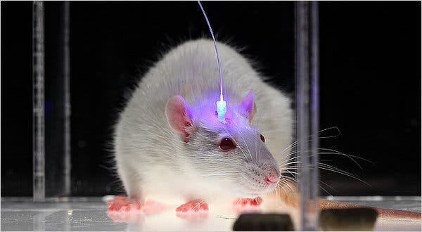
Optogenetics is an innovative biological method that uses light to regulate the way neurons and other cells function in living tissue, combining optical techniques with genetic engineering. It’s a relatively new and emerging technology used in neuroscience, allowing researchers to control individual neurons and study neural circuits across the brain in detail. Along with its applications in understanding brain structure and activity in greater detail, optogenetics shows tremendous promise in treating a variety of neurological disorders in the near future such as epilepsy and even blindness.
Key Points about Optogenetics:
Spatial and temporal resolution are both GREAT
What’s needed to be done: tweak the genes, alter neural activity, use light sensitive proteins, and a fibre optic cable that opens the ion channels when the light is turned on.
…Manipulate brain activity, then what? Behaviour!
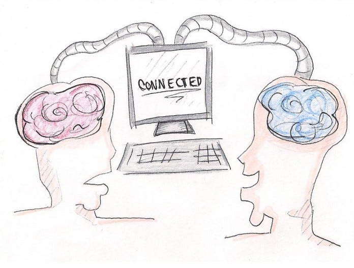
Groundbreaking Brain-to-Brain Study at the University of Washington using EEG + TMS
Using a combination of the internet, neuroimaging and neurostimulation technologies, Rajesh Rao sent a neural signal by his imagination across the UW campus to his colleague Andrea Stocco and stimulated Stocco’s motor cortex. With this study, they seem to have achieved the first noninvasive human-to-human brain interface ever developed.
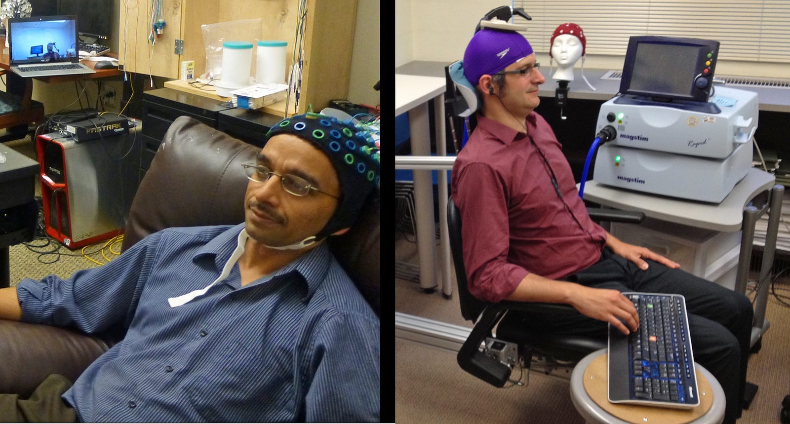
At the University of Washington in Seattle, a “Brain-to-Brain” study was conducted by its Institute for Learning & Brain Sciences (I-LABS) that used a combination of TMS and EEG in which one person’s thoughts control another’s body:
EEG cap → Participant #1, Rajesh Rao
TMS cap → Participant #2, Andrea Stocco*
*Dr. Stocco put a coil with a rapidly changing electric current over a rapidly changing magnetic field on his scalp which induced small electric currents within his brain.
Well you may be wondering, how were these innovative researchers able to achieve this real-life telepathy? The picture and procedural explanation below provides a glimpse into its rather complex setup and the electromagnetic coil experiment that was used to make such a feat possible.

In the experiment, as briefly summarized above, Dr. Rao wears an electroencephalography (EEG) cap, while Dr. Stocco sits in a chair in a separate building across the UW campus with an electromagnetic coil placed above his head. The EEG cap that Dr. Rao was wearing was used to detect his brain activity and send that info to a computer, while the electromagnetic coil was placed directly above Dr. Stocco’s left motor cortex to stimulate activity in that area of his brain (which controls movements on the right side of the body). Now you might ask yourself, where is the connection here considering that they’re in two different buildings across University of Washington’s campus? The connection was actually facilitated via the internet — carrying specific information from the motor area of one brain directly to the motor cortex of another brain via TMS. Specifically, Dr. Rao engaged in a mental video game where he had to shoot targets — something like Call of Duty. Now when he wanted to fire the trigger, he imagined moving his right arm to click the cursor and shoot at a target in the game.
Following this, almost instantaneously, Dr. Stocco — who was not paying any particular attention to the game and had no idea what was going on — started clicking his space bar with his right index finger. Wait what?? Are you telling me that Rajesh’s imagination (remember, he only imagined moving his right arm to fire, he didn’t actually move it) controlled Dr. Stocco’s body? Yes, that’s exactly the case. The reason this happened was that the electromagnetic coil above Dr. Stocco’s cranium was placed in a unique position that caused what he himself described as a “nervous twitch”, which triggered the left motor cortex of his brain to fire a command that moved his right index finger.
It’s safe to say that with a some internet, EEG, and TMS, telepathy was attained with this experiment — yeah I know, it all seems to be straight out of a sci-fi movie! And while the tremendous implications from such a level of brain-to-brain interaction in humans is still novel, the potential applications of such a study is limitless.
As Andrea Stocco mentioned following the study, “years from now the technology could be used, for example, by someone on the ground to help a flight attendant or passenger land an airplane if the pilot becomes incapacitated. Or a person with disabilities could communicate his or her wish, say, for food or water. The brain signals from one person to another would work even if they didn’t speak the same language.” → which speaks to the universality of such a technology.
Andrea Stocco’s wife and research partner Chantel Prat — also a Professor at the University of Washington and author of The Neuroscience of You — beautifully summed up this incredible study by saying, “We plugged a brain into the most complex computer anyone has ever studied, and that is another brain.”
Watch this video to learn more about the experiment (uwneuralsystems on YT):
Neuroimaging and neurostimulation techniques have huge potential, but they all have important limitations that neuroscientists and physicians must be aware of. There are always trade-offs between spatial and temporal resolution regardless of what method is used, and data can be difficult to interpret — even to specialists — as oftentimes they are correlational. But that’s the beauty of it all: each technique has its unique limitations, and that’s why there’s room for multiple. And as we learned from the UW Brain-to-Brain Interface experiment, a combination of these methods can sometimes produce something magical.