The Most Complex Neuroscience Experiment Ever Done
The neuroscientific equivalent of the Human Genome Project
This research breakthrough that was published just this week after almost a decade of experimentation will blow your mind.
Francis Crick, the co-discoverer of the structure of DNA, said this just less than half a century ago: “It is no use asking for the impossible, such as, say, the exact wiring diagram for a cubic millimeter of brain tissue and the way all its neurons are firing.”
Well, that was proven wrong on Wednesday.
An international team of 150 scientists and researchers from 22 institutions including the Allen Institute, Princeton University, Stanford University and the Baylor College of Medicine have done just that, creating the most accurate and complex reconstruction to date of not just the mouse visual cortex—but mammalian brain tissue as a whole—as part of a 9-year project called Machine Intelligence from Cortical Networks (MICrONS). Through this innovative project, researchers aimed to capture in unprecedented detail the structural wiring and neuronal activity of this part of the mouse visual cortex, including how neurons connect and communicate on a micro scale to make perception possible.
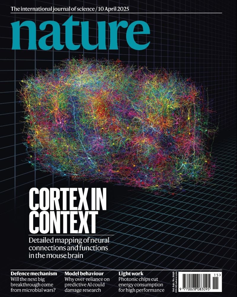
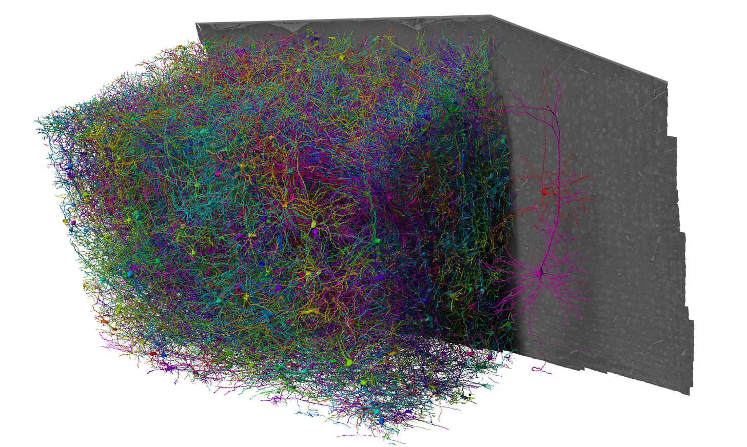
The beautiful image you’re looking at above is the final 3D reconstruction of the cubic millimetre of brain tissue that was analyzed. It is through these interlacing tree-like structures that seem to connect in no particular pattern that mice are able to perceive sight.
A cubic millimetre may seem very little. But believe it or not, within this tiny space the size of a grain of sand are hundreds of billions of connections, hundreds of thousands of nerve cells, and multiple kilometres of axons.
To give you more context, here are some numbers representing exactly what was mapped:
200,000 neurons + neuroglia
523 million synapses
4 kilometres of axons
In-vivo functional data gathered from 75,000 neurons
1.6 petabytes (1.6 million gigabytes) of data used
Truly groundbreaking science.
How did they do it?
The beauty of this research lies in the collaborative nature of it, taking place primarily in multiple world-class institutions and state of the art neuroscience labs across the United States where scientists each worked on a different part of the project, building on the work of their peers.
At Baylor College of Medicine in Houston and Stanford University in California, researchers in Andreas Tolias’s lab gathered all the functional data, using calcium imaging to record the electrical activity of tens of thousands of neurons. To do this, the researchers used a visual stimulating video to stimulate the mouse’s visual cortex and recorded the resulting brain activity from individual neurons in the cortex.
Meanwhile at the Allen Institute for Brain Science in Seattle, scientists used electron microscopy to take a whopping 95 million high-resolution images of that same section of the mouse visual cortex that was studied by researchers at Baylor. With these images, they could precisely see and analyze the neural communication taking place across different networks in the cortex in response to the visual stimulus.
Then in New Jersey, scientists at Princeton University working in Sebastien Seung’s Lab positioned these 95 million high-res images together to produce a 3D reconstruction that revealed this part of the mouse visual cortex and its intricate connections in ridiculous detail.
What’s fascinating is that from the extensive electrical activity data provided by researchers at Baylor, we can infer the intricate neuroanatomy and neurophysiology in the MICrONS wiring diagram. This offers deep insight into both the language and logic that neurons in the brain use to communicate with each other—in other words, the algorithms of the cerebral cortex. → more about the implications of this in the next section
“MICrONS will be remembered as the beginning of a digital transformation of neuroscience. In this new era, computational neuroscience is moving from the fringes to the center of our discipline, and connectomes are becoming the foundation.”
— Dr. Sebastian Seung, Ph.D., Princeton Neuroscience Institute

Getting into some of the more technical details of how all this was done...here’s a quick breakdown:
Within that ‘grain of sand’, functional imaging data of about 75,000 pyramidal neurons was produced, recording single cell responses to a variety of visual stimuli.
As mentioned before, the same area of brain tissue was imaged with high-res electron microscopy from the team at Allen. But then what? These images were processed through a reconstruction pipeline driven by machine learning (ML) that ultimately mapped out a massive anatomical connectome in unparalleled detail—in fact, the largest multi-modal connectomics dataset generated in history by minimum dimension, number of cells (both neurons and neuroglia), and the number of synaptic connections analyzed.
Ridiculous.
What are the broader goals of all this?
Advancing machine learning technology
MICrONS was created with an ambitious goal to revolutionize ML by reverse-engineering complex algorithms within the brain. Mapping the functions and connectivity of diverse cortical circuits with highly advanced imaging technologies is creating new paths for researchers of the MICrONS program to directly use the brain to better understand how ML works, researching the computational inputs and outputs of cortical functions to advance future ML algorithms.
For more precise treatment of neurological pathologies
The innovative research from this experiment has revealed some deep insights into the anatomy and physiology within this region of the mouse visual cortex, which plays a crucial role in brain health and is disrupted commonly by neurological conditions such as Alzheimer’s and autism. By achieving such an incredibly accurate and complex reconstruction of this piece of mammalian brain tissue through the MICrONS experiment, the neuroscience community has taken a HUGE leap forward in revolutionizing our understanding of such neuropsychiatric diseases, the changes they cause in different parts of the brain and ultimately how we can treat them.
Now, with the most precise representation of a connectome ever produced, we can visualize in three dimensions how neurons are connected and how they communicate in this space. Meanwhile, the expansive multimodal datasets from this experiment allow us to comprehensively understand how these neurons communicate from a statistical perspective by analyzing the functional data.
And hey, when it comes to creating reconstructions of this scale of the brain, it’s not easy. Let’s not take for granted how far we’ve come to achieve such a feat. These researchers had to digitally untangle tens of thousands of neurons, literally trace out each neuron’s distinct system of branches, and once that was done did something even more complicated: they reconstructed them one after the other into an extensive network consisting of 500 billion+ connections.
Of course, this was all permitted with the help of recent rapid advances in ML and AI, which I don’t think is something Crick would’ve predicted 46 years ago.

What’s best: this is open data.
You can access the MICrONS Explorer and Allen Institute on Github to explore every bit of data from this multi-year experiment—a powerful research tool that scientists all around the world can freely use in their quests to discover novel treatments for neurological diseases, among other things.
Beautiful.
Now here are some really cool additional tools you can use to analyze and visualize the data on your own, also equipped with instructions on how to query and download portions of the dataset using Python:
For analyzing the anatomical data based on reconstruction of the mouse visual cortex: MICrONS Programmatic Tutorials
For analyzing functional data (you can query functional recordings of neural responses to visual stimuli here): MICrONS Neural Data Access (NDA) repository
I’m a huge fan of open data science, and seeing that these incredible researchers of such a groundbreaking experiment have published all their data on the internet is amazing. Wow. The MICrONS project really built the largest existing dataset of neuroanatomical and neurophysiological data from the mammalian brain and made it open for the public to access. A huge W for science.
Bravo to all of these scientists! The work they’ve done here has revolutionized neuroscience and will reverberate for years to come, serving as the basis for many future experiments that will attempt to do even more.
Innovations like this, with scientists coming together to achieve what was once thought to be impossible, makes me incredibly glad that I study neuroscience in such an exciting day and age.


Thats so cool!